
Early diagnosis and timely Retinal Detachment Treatment can help save vision. Learn about the symptoms, causes, and advanced Retinal Detachment Treatment options in detail.
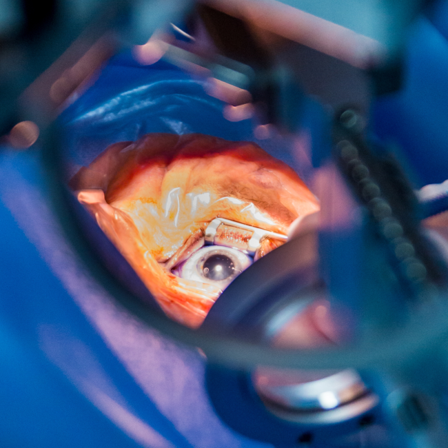
Retinal Detachment is a medical emergency that requires prompt intervention. The retina converts light into signals that travel to the brain, enabling vision. Once detached, the retina cannot function normally, leading to sudden visual changes. Immediate consultation with a Retina Specialist is necessary.
Retinal detachment is a serious eye condition where the retina — the thin layer of tissue at the back of the eye — pulls away from its normal position. This separation prevents the retina from receiving oxygen and nutrients, which can quickly lead to vision loss if not treated immediately.
Common warning signs include sudden flashes of light, a sudden increase in floaters, blurred vision, loss of peripheral vision, and the sensation of a dark curtain moving across your field of vision.
Aging, severe myopia (high minus power), eye trauma, diabetic retinopathy, previous eye surgery, and family history can increase the risk of retinal detachment.
Diagnosis typically involves a dilated eye examination, retinal imaging, OCT scans, and ultrasound evaluation in cases where the retina is not clearly visible.
Treatment may include laser photocoagulation, cryopexy, pneumatic retinopexy, scleral buckle surgery, or vitrectomy depending on the severity and type of detachment.
Recovery depends on how early the condition is treated. Regular eye check-ups, controlling diabetes, and protecting the eyes from trauma help reduce risk.
If you notice sudden flashes, a curtain-like shadow over vision, or sudden loss of sight, consult an eye hospital
immediately. Delaying treatment may result in permanent vision loss.
Eye Examination
Eye Examination
Surgical Procedure
Post-Op Recovery
Retinal detachment happens when the retina, the thin light-sensing layer at the back of the eye, pulls away from the tissue that feeds it. Once it separates, it stops working properly and vision can be lost quickly. It’s a medical emergency, so it needs to be seen by a retina specialist as soon as possible.
Most people notice one or more of these: sudden flashes of light, a sudden shower of new floaters, blurred vision, or a dark shadow creeping in from the side. Some describe it as a curtain being drawn across their sight. It’s usually painless, which is why people sometimes wait too long.
Yes. The longer the retina stays detached, the greater the chance of permanent vision loss. If you notice sudden flashes, a burst of floaters or a shadow across your vision, go to an eye hospital the same day. Don’t wait to see if it settles.
It most often starts with a tear or hole in the retina, which lets fluid slip underneath it. This becomes more likely with age, as the jelly inside the eye (the vitreous) shrinks and tugs on the retina. Other causes include severe short-sightedness, eye injury, previous eye surgery such as cataract surgery, diabetic eye disease, and a family history of detachment.
People with high myopia (a strong minus power) are at higher risk, as are those who’ve had eye trauma or eye surgery, or who have had a detachment in the other eye. Diabetes, lattice degeneration (thin patches in the retina) and a family history also raise the risk. Having a risk factor doesn’t mean you’ll get one, but regular retina checks are sensible.
A retina specialist examines the back of your eye after dilating the pupil with drops. Scans such as OCT or retinal imaging may be used, and an ultrasound is added if the view is blocked, for example by bleeding inside the eye. The whole check is painless and usually quick.
A fully detached retina can’t be fixed with medicines or eye drops. It needs a procedure. Small retinal tears caught early can sometimes be sealed in the clinic with laser treatment or freezing (cryopexy) before they progress to a detachment.
The main options are laser photocoagulation or cryopexy to seal tears, pneumatic retinopexy (a gas bubble placed in the eye), scleral buckle surgery, and vitrectomy. Which one is used depends on where the tear is, how far the detachment has spread, and the condition of your eye. Your surgeon will explain why they’ve recommended a particular approach.
In vitrectomy, the surgeon removes the vitreous gel from inside the eye and seals the retina, usually leaving a gas bubble or silicone oil to hold it in place. In scleral buckle surgery, a small silicone band is stitched to the outside wall of the eye to push it towards the retina and close the break. Vitrectomy is often chosen for older patients and complex cases, and buckling is often used in younger patients.
In published studies, about 85 to 90 percent of retinas reattach with a single operation, and the number rises further if a second procedure is needed. Results are better when the macula, the central part of the retina, is still attached at the time of surgery. How much vision comes back depends mostly on how early the treatment was done.
Not during the operation. It’s done under local or general anaesthesia, depending on the procedure, and you won’t feel it. Afterwards the eye may be sore, red and swollen for a few days, which usually responds well to medicines your doctor prescribes.
Early healing takes a few weeks, but full visual recovery can take a few months. If a gas bubble was used, your vision will be very blurry at first and improve as it shrinks. You may need to keep your head in a certain position for several days, and you must avoid flying and high altitudes until the gas has gone. Your doctor will tell you exactly what to do.
It depends on how long the retina was detached and whether the central part was involved. Some people regain good sight, while others are left with some blurring or distortion even after a successful repair. Early treatment gives the best chance, which is why delay matters so much.
Yes, in some cases. Most redetachments happen in the first few months after surgery, so follow-up visits are important. If it returns, it can usually be treated with another procedure. You’re also at some risk in the other eye, so regular check-ups matter.
Not always, but you can lower your risk and catch problems early. Get regular dilated eye exams if you’re at risk, control diabetes, wear eye protection during sports or work with hazards, and see a doctor promptly for new flashes or floaters. Retinal tears found early can often be treated before they cause a detachment.
Retinal detachment is a sudden mechanical problem where the retina separates from the back of the eye, and it needs urgent surgery. Retinitis pigmentosa is an inherited condition where the retinal cells slowly break down over many years, usually starting with night vision and side vision loss. They’re different conditions with different treatments, so a retina specialist can tell you which one you’re dealing with.
The cost varies with the type of surgery, whether gas or silicone oil is used, the hospital and how complex the case is. Because it’s urgent, it’s wise to ask upfront what’s included, such as tests, medicines, any second procedure and follow-up visits. If you have health insurance, check your cover with your insurer or the hospital’s billing team.

Peason Center for Ophthalmology provides expert Eye Care, advanced diagnostics, and treatments for vision problems.
Hyderabad